| The scanning electron microscope (Philips XL 30 LaB 6, 30kV, see below) is fitted to the image display with a secondary electron detector (SE detector, image REM2). The preferred usage is to illustrate topographycal images (eg fracture surfaces), and a backscatter electron detector (BSE detector, image REM2), etc. to study metallic flat polished specimens. 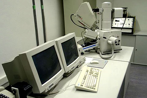
For the measurement of grain orientation of crystalline metallic materials for the EBSD method, a measuring device with appropriate software (OIM) is available. 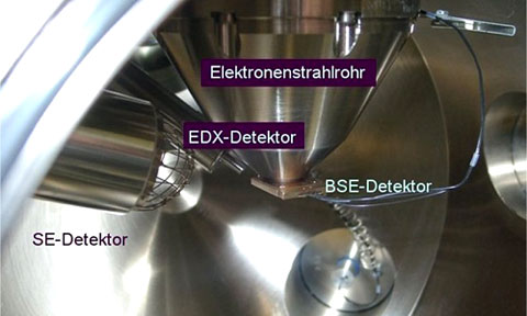
Moreover, the SEM also includes a device (EDX detector, see picture above) and the associated analysis software for the micro-area analysis, which allows qualitative and quantitative information about the local composition. In addition, this software allows the recording of elemental distribution images. |